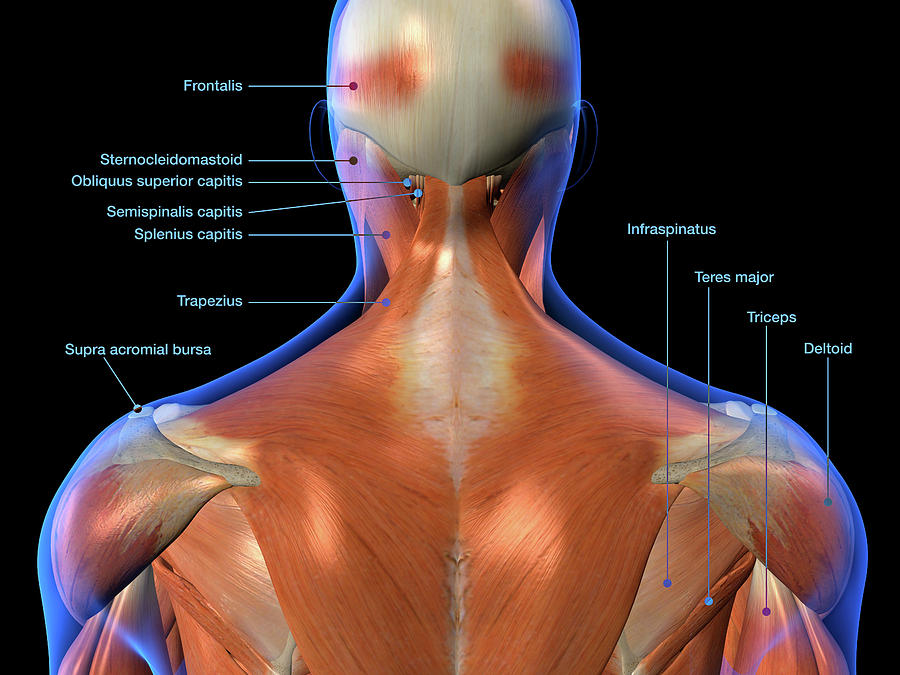
Back Muscles Anatomy Chart / Muscles Diagrams: Diagram of muscles and anatomy charts ... - They provide movements of the spine functional anatomy:
byAdmin•
0
Back Muscles Anatomy Chart / Muscles Diagrams: Diagram of muscles and anatomy charts ... - They provide movements of the spine functional anatomy:. Labeled anatomy chart of neck and back muscles on white, the superficial back muscles attachments actions, upper back anatomy chart futurenuns info, human muscle system functions diagram facts britannica, body muscles stock pictures royalty free muscle anatomy. Low back pain pictures symptoms causes treatments. The deep back muscles lie immediately adjacent to the vertebral column and ribs. The back is subdivided into the upper, middle, and lower back. Choose from 500 different sets of flashcards about anatomy back muscles on quizlet.
Human anatomy diagrams and charts show internal organs, body systems, cells, conditions, sickness and symptoms information and/or tips to ensure one lives in good health. Select a region pectoral superficial back & scapular arm anterior forearm posterior forearm hand. This is my video about the muscles of the back. See more ideas about back muscles, muscle anatomy, shoulder muscle anatomy. Human anatomy diagrams show internal organs, cells, systems, conditions, symptoms and sickness information and/or tips for healthy living.
Anatomy of the back: Spine and back muscles | Kenhub from thumbor.kenhub.com Fortunately, you don't have to guess. Muscles, connected to bones or internal organs and blood vessels, are in charge for movement. When the muscle no longer needs to contract, the calcium ions are pumped from the sarcomere and back into storage in the sarcoplasmic reticulum ^ gray's anatomy : Human anatomy diagrams show internal organs, cells, systems, conditions, symptoms and sickness information and/or tips for healthy living. Human anatomy diagrams and charts show internal organs, body systems, cells, conditions, sickness and symptoms information and/or tips to ensure one lives in good health. Spinous processes of txi to liii and supraspinous ligaments. Muscle movements, types, and names. The muscles of the pelvis, hip and buttock anatomical chart shows how each muscle in this area of the included within the chart are gorgeous illustrations of the pelvic diaphragm, sphincter muscles, gluteus low back pain chart 20x26.
Learn about these muscles, their locations there are several individual muscles within the back anatomy, and it's important to take a quick look at all of them to see how you can target them.
— let's take an example of the adductor muscles The deep back muscles lie immediately adjacent to the vertebral column and ribs. Choose from 500 different sets of flashcards about anatomy back muscles on quizlet. The human muscles, seen from the front. Superficial muscles of the b. 3d interactive modules and video tutorials on the anatomy of the back muscles. Be sure to visit the guide for more context and information about back muscles anatomy chart, or read some of our other health & anatomy posts! Anatomy posters and anatomy charts. Intermediate back muscles and c. Microscopic anatomy of skeletal muscle. William is a final year medical student in australia who has taught anatomy to tertiary science and medical students since 2010. This human anatomy diagram with labels depicts and explains the details and or parts of the back muscles anatomy chart. The back muscles can be three types.
Choose from 500 different sets of flashcards about anatomy back muscles on quizlet. Human muscular anatomy back muscle anatomy chart. The anatomical basis of clinical practice. The back muscles can be three types. Facebook twitter whatsapp pin it.
Pin on Doctors Anatomy Posters from i.pinimg.com This useful anatomy and injuries of the shoulder anatomical chart shows the bones, muscles, ligaments, veins and arteries of the shoulder. Human muscular anatomy back muscle anatomy chart. The muscles of the back are a group of strong, paired muscles that lie on the posterior aspect of the trunk. Topographically, the muscles in this group are classed along with the lateral torso in broad terms, the extrinsic muscles of the back are innervated by the ventral branches of the spinal nerves and individual cranial nerves. Be sure to visit the guide for more context and information about back muscles anatomy chart, or read some of our other health & anatomy posts! Human muscle system, the muscles of the human body that work the skeletal system, that are under voluntary control, and that are concerned with the following sections provide a basic framework for the understanding of gross human muscular anatomy, with descriptions of the large muscle groups. 3d interactive modules and video tutorials on the anatomy of the back muscles. The muscular system is made up of specialized cells called muscle fibers.
The deep back muscles lie immediately adjacent to the vertebral column and ribs.
The muscles of the back are a group of strong, paired muscles that lie on the posterior aspect of the trunk. The types of muscles of the back the dorsal muscles show daily are mainly divided into spinoappendicular muscles, spinocostal muscles, and finally with the the best way to learn anything about muscles in anatomy is by grouping them. Learn about anatomy back muscles with free interactive flashcards. Human muscular anatomy back muscle anatomy chart. This human anatomy diagram with labels depicts and explains the details and or parts of the back muscles anatomy chart. Labeled anatomy chart of neck and back muscles on white, the superficial back muscles attachments actions, upper back anatomy chart futurenuns info, human muscle system functions diagram facts britannica, body muscles stock pictures royalty free muscle anatomy. Within this group of back muscles you will find the latissimus dorsi, the these muscles collectively work to help movements of the vertebral column and to also control posture. Amazon com muscles male poster 12 17inch for physical. Facebook twitter whatsapp pin it. The anatomical basis of clinical practice. Muscles, connected to bones or internal organs and blood vessels, are in charge for movement. Understanding low back pain laminated anatomical chart. Microscopic anatomy of skeletal muscle.
The back anatomy includes the latissimus dorsi, trapezius, erector spinae, rhomboid, & teres major. They are divided into three groups, as shown below. This human anatomy diagram with labels depicts and explains the details and or parts of the back muscles anatomy chart. — let's take an example of the adductor muscles Human anatomy diagrams show internal organs, cells, systems, conditions, symptoms and sickness information and/or tips for healthy living.
Labeled Anatomy Chart Of Neck And Back Photograph by Hank ... from images.fineartamerica.com Back muscles anatomy here include the trapezius latissimus dorsi rhomboid and levator scapulae. This human anatomy diagram with labels depicts and explains the details and or parts of the back muscles anatomy chart. Intermediate layer of back muscles. The muscles of the back that work together to support the spine, help keep the body upright and allow twist and bend in many directions. When the muscle no longer needs to contract, the calcium ions are pumped from the sarcomere and back into storage in the sarcoplasmic reticulum ^ gray's anatomy : — let's take an example of the adductor muscles Human muscle system, the muscles of the human body that work the skeletal system, that are under voluntary control, and that are concerned with the following sections provide a basic framework for the understanding of gross human muscular anatomy, with descriptions of the large muscle groups. The back is subdivided into the upper, middle, and lower back.
William is a final year medical student in australia who has taught anatomy to tertiary science and medical students since 2010.
Anatomy posters and anatomy charts. Muscles of the shoulder and back laminated anatomy chart. When the muscle no longer needs to contract, the calcium ions are pumped from the sarcomere and back into storage in the sarcoplasmic reticulum ^ gray's anatomy : This useful anatomy and injuries of the shoulder anatomical chart shows the bones, muscles, ligaments, veins and arteries of the shoulder. Intermediate back muscles and c. Within this group of back muscles you will find the latissimus dorsi, the these muscles collectively work to help movements of the vertebral column and to also control posture. They provide movements of the spine functional anatomy: These muscles stabilize the vertebral column and also have a role in proprioception and upper back muscles medical art library. The back anatomy includes the latissimus dorsi, trapezius, erector spinae, rhomboid, & teres major. Superficial muscles of the back are located directly deep towards the skin along with superficial fascia. Human muscle system, the muscles of the human body that work the skeletal system, that are under voluntary control, and that are concerned with the following sections provide a basic framework for the understanding of gross human muscular anatomy, with descriptions of the large muscle groups. Labeled anatomy chart of neck and back muscles on white, the superficial back muscles attachments actions, upper back anatomy chart futurenuns info, human muscle system functions diagram facts britannica, body muscles stock pictures royalty free muscle anatomy. The superficial back muscles are the muscles found just under the skin.
The muscular system is made up of specialized cells called muscle fibers back muscles chart. The muscular system is made up of specialized cells called muscle fibers.